Community-acquired Klebsiella pneumoniae – Its Virulence and Antimicrobial Resistance Genes Complement in Participants Presenting for COVID-19 Testing in Lagos State, Nigeria
 , Jacob Itopa Yisau, MSc1, Toun Wuraoluwa Fesobi, MSc1, Muinah Adenike Fowora, PhD1, Tajudeen Akanji Bamidele, PhD1, Toyosi Yekeen Raheem, PhD1, Adenike Shola Aiyedogbon, MSc1, Ibilola Omolopo, MSc1, Abraham Ajayi, PhD1, Olufemi Samuel Amoo, PhD2, Chinonso Ikpo, BMLS1, Grace Abosede Akintunde, MSc1, Bamidele Abiodun Iwalokun, PhD1, Stella Ifeanyi Smith, PhD1, Babatunde Lawal Salako, MBBS, FWACP1

*Corresponding author: Ajoke Olutola Adagbada, Department of Molecular Biology and Biotechnology, Nigerian Institute of Medical Research, Yaba, Lagos, Nigeria. jokejola2002@yahoo.com
-
Received: ,
Accepted: ,
Abstract
Background and Objective
The COVID-19 pandemic brought about significant health, economic, and security challenges during its outbreak in late 2019. Accurate diagnosis and differentiation from other respiratory infections are crucial for effective management. Klebsiella pneumoniae, a known respiratory pathogen, presents symptoms similar to COVID-19, complicating diagnosis and treatment. This study emphasizes the importance of screening for K. pneumoniae in individuals presenting with COVID-19-like symptoms to achieve proper patient management.
Methods
In this cross-sectional study, a total of 269 nasopharyngeal swabs were collected from individuals at a COVID-19 testing center in Yaba, Lagos. Standard laboratory procedures were employed for the isolation of K. pneumoniae using MacConkey and Eosin Methylene Blue agars, characterization was done with specific biochemical tests, and antibiotic susceptibility testing using the disc diffusion method. Polymerase chain reaction was used to detect species-specific, virulence, and antimicrobial resistance genes.
Results
Among the participants, 11.2% tested positive for COVID-19, 7.8% for Klebsiella species, and 0.74% for both. Specifically, 5.24% were positive for the K. pneumoniae-specific gene, PfPr1. Antibiotic susceptibility ranged from 30.4% to 82.6%, with sensitivities to cephalosporins (47.8%), carbapenems (65.3%), and ciprofloxacin (34.8%). Notably, all isolates were resistant to macrolides and glycopeptides. Of the 14 K. pneumoniae isolates, 7 harbored virulence genes: 4 were positive for mrkD (28.6%) and 3 for K2 (21.4%). In addition, 12 isolates carried antimicrobial resistance genes: 10 were positive for blaTEM (71.4%) and 2 for blaSHV (14.3%).
Conclusion and Implication for Translation
The detection of virulence factors and antimicrobial resistance genes, including those conferring resistance to last-resort antibiotics, in community-acquired K. pneumoniae isolates highlights the potential for severe clinical outcomes and increased healthcare burden. The emergence of these multidrug-resistant strains in community settings underscores the critical need for routine surveillance of K. pneumoniae, particularly in the context of coinfections with Severe Acute Respiratory Syndrome Coronavirus-2 and the implementation of comprehensive antimicrobial stewardship programs to mitigate the spread and impact of resistant pathogens. This will lead to improved case management in the event of a new wave of COVID-19 infection.
Keywords
COVID-19
Genes
Klebsiella pneumonia
Resistance
Virulence
INTRODUCTION
The COVID-19 pandemic has significantly impacted global health negatively, in the area of respiratory tract infections, emphasizing the need for its accurate diagnosis and differentiation from other respiratory tract infections caused by bacterial agents. Klebsiella pneumoniae, a Gram-negative bacterium, is an opportunistic pathogen originating from the gastrointestinal tract but capable of causing respiratory infections with symptoms resembling COVID-19.
From earlier studies, 8–12% of hospital-acquired pneumonia occurs in patients due to the use of ventilators, and such pneumonia is caused by K. pneumoniae; however, community-acquired infection with K. pneumoniae accounts for 3–5% and 15% of pneumonia cases in developed and developing countries, respectively.[1]
Co-infection of COVID-19 with other bacterial respiratory infections presents a challenge that complicates treatment. Co-infection of COVID-19 and K. pneumoniae presents an increased risk of morbidity/mortality, especially for hospitalized patients and high-risk individuals. Some authors[2] in a systematic review revealed that co-infected patients in Asia, Europe, and Africa are 23%, 15%, and 43% respectively. However, insufficient data and surveillance hinder effective management and monitoring of such co- infection in Nigeria. K. pneumoniae causes significant disease in immuno- compromised and hospital patients. Pneumonia due to K. pneumoniae is an important cause of morbidity and mortality, especially in hospital settings. Associated symptoms include shortness of breath, fever, sputum production, and pleuritic chest pain. However, the burden of community-acquired infection due to this pathogen and its attendant morbidity has been on the increase in recent years.[3] K. pneumoniae is classified into two pathotypes: Classical and hypervirulent. Hypervirulent strains have extra virulence factors enabling them to cause disease in healthy individuals in community settings; classical is an opportunistic pathogen that mostly causes hospital-acquired infection in the immunocompromised.[4] Lately, hypervirulent serotypes of this pathogen are associated with diseases in immunocompetent and healthy individuals, causing life- threatening community-acquired infections, including pneumonia.[5,6]
This pathogen’s virulence factors include capsular antigen, based on which the organism was classified into 79 serotypes, K1 to K79. K1 and K2 capsular types are common with hypervirulent pathotypes and fimbriae.[5,6] K. pneumoniae contributes to antimicrobial resistance through possession of extended-spectrum beta-lactamase (ESBL) and carbapenemase genes, making it resistant to cephalosporins and beta-lactams. This increases its potential for causing severe morbidity and mortality.[5,6]
During the COVID-19 pandemic, for the management of patients with respiratory illnesses, asymptomatic treatment in many instances would commence before the etiology is determined. This exerts further selective genetic pressure on the pathogen, causing the acquisition of greater pathogenic potential. Therefore, it is essential that, in addition, screening is done for bacterial infections to achieve optimal management.
This study investigated K. pneumoniae in participants presenting for COVID-19 testing at a national reference center in Lagos, Nigeria. The aim was to determine the concurrence of K. pneumoniae with COVID-19, the genetic basis of its antimicrobial resistance, and virulence determinants. The objectives were to evaluate the prevalence of K. pneumoniae, determine the prevalence of COVID-19 and K. pneumoniae co-infection in participants reporting for COVID-19 testing, to determine the antibiotic resistance profiles of the isolates, and to assess their virulence and antimicrobial gene complement.
METHODS
Study Setting and Duration
This cross-sectional study was carried out on drive-in and walk-through COVID-19 testing facilities in the Nigerian Institute of Medical Research (NIMR), Lagos, Nigeria. The NIMR is a leading medical research institute located in Yaba, an urban area in the commercial capital (Lagos) of Nigeria. Lagos is the most populous city in Nigeria, with approximately 24 million inhabitants (https://lagosstate.gov. ng/about-lagos). During the COVID-19 pandemic, the drive- in and walk-through COVID-19 testing center in the NIMR received more than 50,000 people, making it one of the largest in the country. Sample collection for this study took place between June and October 2022 during the COVID-19 pandemic.
Ethical Approval/Recruitment of Participants and Collection of Data
Ethical approval (IRB/21/069) was obtained from NIMR Institutional Review Board (NIMR IRB). Using convenience sampling,[7] a total of 269 participants who gave informed consent were recruited for the study. The demographic characteristics (age and sex) and other data of study participants were collected with a structured questionnaire. Individuals who did not give their consent to participate in the study were excluded. There were no other exclusion criteria.
Collection of Nasopharyngeal Swabs
Following the guidelines of the Nigeria Center for Disease Control,[8] 269 nasopharyngeal swabs were collected from participants under strict sterile conditions in duplicates into both bacterial transport medium (designed to inhibit viral particles: Skim milk-tryptone-glucose-glycerine) and viral transport medium (designed to suppress bacterial growth). These were kept on an ice pack following standard operating procedures for COVID-19 infection, and thereafter transported to the laboratory for further laboratory testing.
Isolation of Bacteria
Nasopharyngeal swabs were inoculated on MacConkey agar (serving as both primary isolation and differential medium) and incubated at 37°C for 24 hour (h) using standard microbiological procedures.
Suspected K. pneumoniae colonies were then subcultured on Eosin Methylene Blue agar and incubated at 37°C for 24 h. The characteristic purple, dark-centered, mucoid colonies were presumptive of K. pneumoniae. Suspected colonies were characterized and presumptively identified based on morphological characteristics such as color, shape, Gram staining, and biochemical tests, including sugar fermentation, citrate utilization, and the string test was done as previously described by Khan et al.[9] Presumptively identified K. pneumoniae were stored at −80°C for further analysis.
Antimicrobial Susceptibility Testing
The K. pneumoniae isolates were tested for antibiotic susceptibility following EUCAST v13.0 clinical breakpoints and guidelines for enterobacterales.[10]
With American Type Culture Collection (ATCC 13883) K. pneumoniae strain as the quality control strain, Kirby–Bauer agar disc diffusion method[11] was deployed to determine the antibiotic susceptibility pattern of isolates, using the following class of antibiotics (Oxoid): Cephalosporin (Ceftazidime [10 µg]), Ceftriazone (30 µg), Quinolone (Ciprofloxacin CIP [5 µg]), Glycopeptide (Vancomycin [5 µg]), Macrolide (Erythromycin E [15 µg]), Azithromycin (15 µg), Carbapenem (Imipenem IMI [10 µg]), Meropenem (10 µg), Penicillin (Augmentin [30 µg]), and Nitrofurantoin (300 µg).
A suspension of pure bacterial colonies was made in normal saline up to the McFarland standard 0.5. The sterile swab stick was used to make lawns of suspension on sterile Mueller– Hinton agar plates, after which they were left to dry. The antibiotic discs were then aseptically placed on top, and the plates incubated at 37°C for 24 h. Zones of inhibition were measured with the aid of a meter rule in millimeters and interpreted as sensitive (S), intermediate (I), and resistant (R). Breakpoints for susceptibility and resistance were determined using the EUCAST Breakpoint Tables (Version 13.0).
Genomic Deoxyribonucleic Acid (DNA) Extraction
DNA extraction for the detection of species-specific, virulence, and antimicrobial resistance genes was done using a modification of the method described by Goudoudaki et al.[12] Cells from an overnight culture were harvested into 1000 µL of sterile distilled water in a 1.5 mL Eppendorf tube and vortexed to mix. This was spun at 10,000 revolutions/ min (rpm) for 5 min, and the supernatant was discarded. The cell pellets were resuspended in 1000 µL of sterile distilled water, vortexed, and spun at the previously mentioned speed and for the same duration. The supernatant was again discarded, and the pellets resuspended in 200 µL of distilled water, vortexed, and incubated at 100°C for 5 min, after which it was transferred to an ice block and incubated for 20 min. This was spun again at 10,000 rpm, the supernatant was transferred into a microcentrifuge tube, and stored at −20°C for further use.
Genomic DNA Quantiflcation and Purity
The concentration and purity of extracted DNA were determined using the NanoDrp oneC spectrophotometer (Thermo Fisher Scientific, South Africa Intertek 58595). Upon validation of satisfactory yield and purity, they were deployed for gene detection.
Identiflcation of K. pneumoniae and Molecular Detection of Virulence Determinants and Antibiotic Resistance Genes
Polymerase chain reaction (PCR), using SimpliAmp Thermal Cycler (Thermo Fisher Scientific), was deployed for the detection of antibiotic resistance genes (blaTEM and blaSHV), virulence genes (K2 and mrkD), and the detection of Pf/Pr1 gene for the molecular identification of K. pneumoniae using sets of primers listed in Table 1. A 20 µL PCR reaction mixture was made up of 2.5 µL template DNA, 4 µL of 2X Master Mix, 0.5 µL each of the forward and reverse primers, and 12.5 µL of nuclease- free PCR water to make a final volume of 20 µl for each reaction tube. The cycling parameters, which consisted of initial denaturation at 95°C for 5 min, denaturation at 95°C for 30 seconds (s), annealing at 50°C for 30 s, extension at 72°C for 1 minutes (min) and final extension at 72°C for 10 min were the same for all the primers except for blaTEM, for which the annealing condition was 45°C for 30 s. PCR products were separated alongside a 100 bp ladder by electrophoresis on a 2% agarose gel at 80V for 35 min and visualized under a UV transilluminator.
Data Analysis
Data analysis was done using R Statistical Programming Software version 4.3.1 to analyze the participants’ demographic data and laboratory results.
This cross-sectional study was conducted in compliance with the Strengthening the Reporting of Observational Studies in Epidemiology guidelines.[13]
RESULTS
Demographic Distribution of the Participants
A total of 269 participants were enrolled in the study. The minimum and maximum ages were 3 and 80, respectively, with a mean age of 33.5 (±13.4) years. The sex distribution of the participants consists of 117 (43.5%) females, 131 (48.7%) males, while 21 (7.8%) did not register their sex. Among the 32 positive COVID-19 results, 11 males and 21 females were positive.
Infections among Study Participants
As shown in Figure 1, out of the 269 nasopharyngeal samples collected, 32 (11.2%) were positive for COVID-19, while 21 (7.8%) were positive for K. pneumoniae. However, molecular identification indicated that 14 (5.2%) were K. pneumoniae. Two (0.74%) of the samples were positive for both COVID-19 and K. pneumoniae (co-infection). Therefore, the prevalence of COVID-19, K. pneumoniae infection, and COVID-19/K. pneumoniae co-infection was 11.2%, 7.8%, and 0.74%, respectively.
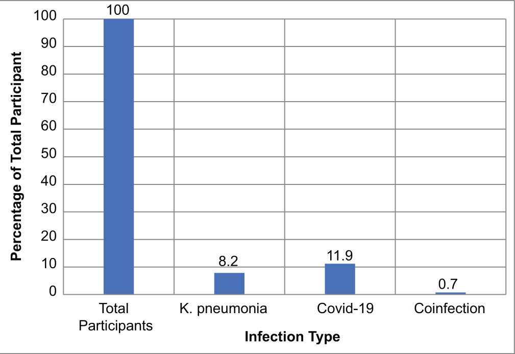
- Infections among study participants.
Antimicrobial Resistance Pattern of the K. pneumoniae Isolates
The antimicrobial resistance pattern of the isolates, as shown in Figure 2, showed a general resistance to the macrolides and glycopeptides (100%), but susceptibility ranging from 30.4% to 82.6% to the other antimicrobials: 47.8% of the isolates were susceptible to cephalosporins, 65.25% of isolates were susceptible to carbapenems while 34.8% of the isolates were susceptible to ciprofloxacin.
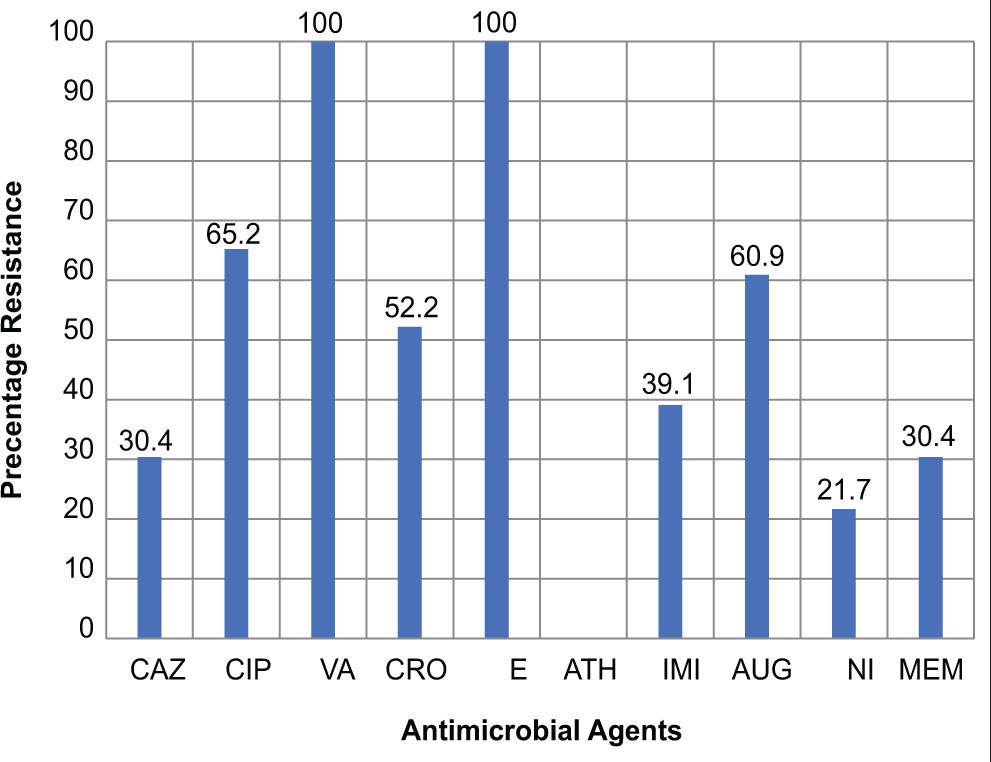
- Antimicrobial resistance pattern. CAZ: Ceftazidime, CIP: Ciprofloxacin, VA: Vancomycin, CRO: Ceftriaxone, E: Erythromycin, ATH: Azithromycin, IMI: Imipenem, AUG: Augmentin, NI: : Nitrofuratoin, MEM: Meropenem
Figure 3 shows the detection of K. pneumoniae using the species-specific gene PfPr1, while Figure 4 shows the Gel picture showing blaTEM gene PCR products from K. pneumoniae.
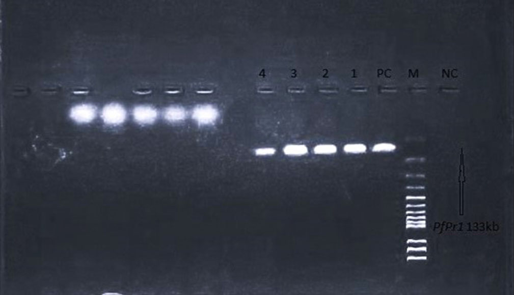
- Gel picture showing detection of Klebsiella pneumoniae using the species-specific gene PfPr1. Lane 1: Negative control, Lane 2: Marker, Lane 3: Positive control, Lanes 4-6: Sample results.
Genes, Sequence of Primers, and Size of the Fragments
Table 1[14-16] displays the genes and primer sequences used in this study.
As shows in Table 2, out of the 14 K. pneumoniae isolates, 4 were positive for mrkD (28.6%) and 3 were positive for K2 (21.4%); also, 10 were positive for blaTEM (71.4%) and 2 were positive for blaSHV (14.3%). Virulence genes and antimicrobial resistance genes combinations were also observed among the isolates. One of the isolates carried all four genes studied, one harbored 3 different genes, whereas 4 harbored 2 different genes.
| Target gene | Primer name | Primers sequence (5’ ‑3’) | Amplicon size (bp) | References |
| 16S−23S rDNA internal transcribed spacer | Pf/Pr1 | 5’-ATT TGA AGA GGT TGC AAA CGA T-3’ 5’-TTC ACT CTG AAG TTT TCT TGT GTT C-3’ |
133 | [14] |
| Adhesin type 3 fimbriae | mrkD | F_AAGCTATCGCTGTACTTCCGGCA R_GGCGTTGGCGCTCAGATAGG | 340 | [14] |
| Capsular serotype K2 and hypermucoviscosity phenotype | K2 | F_CAACCATGGTGGTCGATTAG R_TGGTAGCCATATCCCTTTGG | 531 | [15] |
| ESBL encoding gene | blaTEM | F–ATGAGTATTCAACATTTCCG R–CCAATGCTTATTCAGTGAGG | 858 | [16] |
| ESBL encoding gene | blaSHV | F–TTATCTCCCTGTTAGCCACC R–GATTTGCTGATTTCGCTCGG | 797 | [16] |
ESBL: Extended-spectrum beta-lactamase
| Gene combination | Number of positive isolates | Percentage of positive isolates |
| Pf/Pr1 | 14 | 100.0 |
| blaTEM | 10 | 71.4 |
| blaSHV | 2 | 14.3 |
| K2 | 3 | 21.4 |
| mrkD | 5 | 35.7 |
| mrkD/K2 | 1 | 7.1 |
| mrkD/blaTEM | 2 | 14.3 |
| K2/blaTEM | 1 | 7.1 |
| K2/blaTEM/blaSHV | 1 | 7.1 |
| mrkD/K2/blaTEM/blaSHV | 1 | 7.1 |
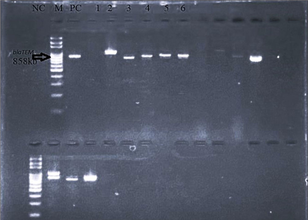
- Gel picture showing blaTEM gene PCR products from Klebsiella pneumonia. Lane 1: Negative control, Lane 2: Marker, Lane 3: Positive control, Lanes 4-7: sample results.
DISCUSSION
K. pneumoniae causes significant morbidity and mortality in the infirm and immunocompromised patients in hospital settings, and increasingly in healthy humans in community settings. Capsular types associated with the hypervirulent K. pneumoniae include the K1 and K2. Aside from their virulence, K. pneumoniae also carries a number of resistance genes, including ESBL and carbapenemase, which render treatment options limited, expensive, or complex.
This study showed a prevalence of 11.9%, 8.2% and 0.74% for COVID-19, K. pneumoniae, and COVID-19/K. pneumoniae coinfection. This has been demonstrated in some earlier studies.
The prevalence of COVID-19 and Klebsiella spp. coinfection in Africa, as reported in a systematic review/meta-analysis report, is 43%, far higher than that of Asia (23%) and Europe (15%).[2] The prevalence in the report is higher than the one observed in this study, probably because the study covers community and hospital-based studies.
The COVID-19 prevalence of 11.9% in this study was lower than 16.3%[7] and the difference could be because the COVID-19 wave was sloping downwards during this study.
Of 136 patients with community-acquired pneumonia, a study recorded 11.0% K. pneumoniae infections,[17] which is similar to our study’s record of 8.2% while[18] reported that Gram-negative enterobacteria, represented predominantly by K. pneumoniae, were 3.7% of isolates in hospital-acquired pneumonia associated with COVID-19.[18] The prevalence is also significantly lower than 30.5% from a study in Southwestern Nigeria,[19] which was conducted on clinical samples.
They exhibited susceptibility ranging from 30.4% to 82.6% to other antimicrobials. They also exhibited 52.2%, 34.7%, and 65.2% resistance to the cephalosporin, carbapenems, and CIP, respectively.
High resistance of the isolates to third-generation cephalosporins (52.2%) and carbapenem (34.7%), as observed in this study, was also observed in a study in Lagos state by Akinyemi et al.[20] In addition to this, in a similar Lagos state study, isolates showed 27.4% resistance to carbapenems[21] and 61.20%[22] as opposed to IMI with 16.3% resistance, a lower figure recorded in Nasarawa state.[23]
This study also reveals isolates that combine a potent mix of virulence and antimicrobial resistance genes, which is noteworthy because they are obtained from participants from the community setting, raising concern about the severity of the disease they cause and the cost of treatment. The virulence genes observed in our isolates are mrkD and K2. The mrkD gene encodes the mrkD protein, which helps in bacterial adhesion, mediating the binding of the organism to the extracellular matrix, leading to biofilm formation, which prevents penetration of antibiotics during therapy and thus poses treatment difficulty. The K2 is capsular serotype- specific, and it mediates the organism’s evasion of the host immune system.
Among the isolates, 28.6% and 21.4% were positive for the mrkD and K2 genes, respectively. The K2 capsular types exhibit a high virulence phenotype and are predominant in invasive infections; this study reported a prevalence of 21.4% for the K2 gene, which differs markedly from some published data on the gene: 2%[24] and 8%.[25] The prevalence of the mrkD gene among our isolates, while being in a similar range with that reported by Imtiaz et al.,[26] differs markedly from those reported by Ahmadi et al.,[27] Bakhtiari et al.,[28] This difference could be attributed to the fact that in contrast to our isolates are community-acquired, while the reported isolates are hospital-related and would have been subjected to greater selective/evolutionary pressure, and this is further evidenced by the observation in the reports that there were associations found between some of the virulence genes and the antimicrobial resistance genes.[27,28]
The prevalence of the antimicrobial resistance genes in this study is 14.3% for blaSHV and 71.4% for blaTEM. For the blaTEM gene, our data differ from those reported by Latifpour et al.,[29] Ayeni et al.[30] who reported rates of 32%, 47.7%, 16%, 17.3%, respectively, but agrees with that reported by Odewale et al.[19] with a prevalence of 89.5%.[29] Worked on both community-acquired and hospital-based infections.[20] Also reported blaSHV in K. pneumoniae strains in a Lagos- based study, while in a study conducted in Nasarawa state, the percentage occurrence of Carbapenem resistance genes blaKpc and blaVIM was 100% and 33.3% positive, respectively.[31]
Limitations of the Study
The different waves of COVID-19 affected sampling during this study. Recruitment of participants and sample collection for the study took place during the 4th wave of the COVID-19 pandemic in Lagos, Nigeria. Therefore, having no prior knowledge of how long the 4th wave would last, we adopted a convenience sampling method to be able to recruit as many participants as the situation afforded. This may affect the broad generalizability of our findings, and we acknowledge it as a limitation of the study.
Lack of sufficient funding also brought about some limitations. In addition, some of the participants later declined their consent to participate in the study.
The isolates from our study possessed ESBL genes, including blaTEM and blaSHV. Further studies may consider determining the actual prevalence of these genes in K. pneumoniae isolates in Lagos and provide a basis for quantifying the resistance conferred by these genes.
CONCLUSION AND IMPLICATIONS FOR TRANSLATION
Coinfection between COVID-19 and K. pneumoniae significantly affects the clinical severity of patients and poses challenges to diagnosis and treatment strategies.
Prevalence of 11.2%, 7.8% and 0.74% for COVID-19, K. pneumoniae, and COVID-19/K. pneumoniae coinfection was reported in this study. Prevalence of the antimicrobial resistance genes in isolates in this study was 14.3% for blaSHV and 71.4% for blaTEM. Noting that the isolated K. pneumoniae are community- acquired with some of the isolates being resistant to the last-resort antibiotics, and possessing a high pathogenic capacity indicated by the presence of virulence and antimicrobial resistance genes, we suggest that patients presenting for COVID-19 testing should also be screened for presence of coinfection with K. pneumoniae; and we propose the following in the event of a future wave of COVID-19; Screening for K. pneumoniae alongside COVID-19 testing so as to rule out respiratory bacterial infections or co-infection and proper antimicrobial management; surveillance to determine the environmental reservoir, and more community-based studies to shed light on the incidence. This will lead to an improvement in the treatment plan and management options of respiratory illnesses in a COVID-19 outbreak.
Key Messages
(1) In the event of a future COVID-19 outbreak, Klebsiella pneumoniae screening can be included in the clinical diagnosis. (2) Antibacterial drugs should be included in the treatment regimen, especially in fulminant cases of COVID-19 during the outbreak. (3) More research will need to be initiated, especially at the onset of a pandemic, on the impact of bacterial coinfection on the severity of COVID-19.
Acknowledgments
This work was supported by the Nigerian Institute of Medical Research (NIMR) COVID-19 Research Support (Grant Number NMG-CIF-21-0069). This work was also supported by the Kano Independent Research Center Trust. We acknowledge the management for their collaboration with NIMR and for supporting the payment of the manuscript fees.
COMPLIANCE WITH ETHICAL STANDARDS
Conflicts of Interest: There are no conflicts of interest. Financial Disclosure: Nothing to declare. Funding/Support: This work was supported by the Nigerian Institute of Medical Research (NIMR) COVID-19 Research Support (Grant Number NMG-CIF-21-0069). This work was also supported by the Kano Independent Research Center Trust (KIRCT). We acknowledge the management of KIRCT for their collaboration with the Nigerian Institute of Medical Research and for supporting the payment of the manuscript fees. Ethics Approval: This study is approved by the Institutional Review Board at Nigerian Institute of Medical Research (NIMR), number IRB/21/069, dated 2021. Declaration of Patient Consent: The authors certify that they have obtained all appropriate patient consent. Use of Artificial Intelligence (AI)‑Assisted Technology for Manuscript Preparation: The author(s) confirm that there was no use of Artificial Intelligence (AI)-Assisted Technology for assisting in the writing or editing of the manuscript, and no images were manipulated using the AI. Disclaimer: None.
References
- Klebsiella pneumonia. In: StatPearls. Treasure Island, FL: StatPearls Publishing; 2025.
[Cited 2025 Jul 24]. Available from: https://www.ncbi.nlm.nih.gov/books/nbk519004 - [Google Scholar]
- Co-infection of SARS-CoV-2 and Klebsiella pneumoniae: A systematic review and meta-analysis. Diagnostics (Basel). 2024 May 30;14(11):1149.
- [CrossRef] [PubMed] [Google Scholar]
- Klebsiella pneumoniae bacteraemia epidemiology: Resistance profiles and clinical outcome of King Fahad Medical City isolates, Riyadh, Saudi Arabia. BMC Infect Dis. 2023 Sept 05;23(1):579.
- [CrossRef] [PubMed] [Google Scholar]
- Virulence analysis and antibiotic resistance of Klebsiella pneumoniae isolates from hospitalized patients in Poland. Sci Rep. 2023 Mar 17;13(1):4448.
- [CrossRef] [PubMed] [Google Scholar]
- Virulence factors in hypervirulent Klebsiella pneumoniae. Front Microbiol. 2021 Apr 08;12:642484.
- [CrossRef] [PubMed] [Google Scholar]
- Hypervirulent Klebsiella pneumoniae. Clin Microbiol Rev. 2019 May 15;32(3):e00001-19.
- [CrossRef] [PubMed] [Google Scholar]
- Prevalence and presentation of pediatric coronavirus disease 2019 in Lagos, Nigeria. Int J Pediatr. 2021 Oct 06;2021:2185161.
- [CrossRef] [PubMed] [Google Scholar]
- National interim guidelines for clinical management of COVID-19. National interim guidelines for clinical management of COVID-19.
[Cited 2025 Jul 24]. Available from: https://ncdc.gov.ng/themes/common/docs/protocols/177_1584210847.pdf - [Google Scholar]
- Comprehensive analysis of Klebsiella pneumoniae culture identification and antibiogram: Implications for antimicrobial susceptibility patterns from sputum samples in Mardan, Khyber Pakhtunkhwa, Pakistan. Bacteria. 2023 Nov 15;2(4):155-73.
- [CrossRef] [Google Scholar]
- Breakpoint tables for interpretation of MICs and zone diameters Version 13.1, valid from 2023-06-29. Sweden: EUCAST.
European Committee on Antimicrobial Susceptibility Testing. [cited 2024 Dec 02]. Available from: https://www.eucast.org/fileadmin/src/media/pdfs/eucast_files/breakpoint_tables/v_13.1_breakpoint_tables.pdf - [Google Scholar]
- Conventional methods and future trends in antimicrobial susceptibility testing. Saudi J Biol Sci. 2023 Feb 10;30(3):103582.
- [CrossRef] [PubMed] [Google Scholar]
- Fast and sustainable thermo-osmotic DNA extraction protocol for trans-spectrum contingency and field use. Bio Protoc. 2023 Sept 05;13(17):e4796.
- [CrossRef] [PubMed] [Google Scholar]
- The strengthening the reporting of observational studies in epidemiology (STROBE) statement: Guidelines for reporting observational studies. PLoS Med. 2007 Oct 16;4(10):e296.
- [CrossRef] [PubMed] [Google Scholar]
- PCR detection of Klebsiella pneumoniae in infant formula based on 16S-23S internal transcribed spacer. Int J Food Microbiol. 2008 Jul 31;125(3):230-5.
- [CrossRef] [PubMed] [Google Scholar]
- Multiplex PCR for detection of seven virulence factors and K1/ K2 capsular serotypes of Klebsiella pneumoniae. J Clin Microbiol. 2014 Dec;52(12)4377-80.
- [CrossRef] [PubMed] [Google Scholar]
- Prevalence of Klebsiella pneumoniae encoding genes for Ctx-M-1, Tem-1 and Shv-1 extended- spectrum beta lactamases (ESBL) enzymes in clinical specimens. Jundishapur J Microbiol. 2013;6(10):e8256.
- [CrossRef] [Google Scholar]
- The COVID-19 pandemic and the incidence of the non-COVID-19 pneumonia in adults. Front Med (Lausanne). 2021 Nov 11;8:737999.
- [CrossRef] [PubMed] [Google Scholar]
- Etiology of community-acquired and hospital- acquired pneumonia associated with COVID-19. Int J Infect Dis. 2022 Mar;116:S39.
- [CrossRef] [Google Scholar]
- Genotypic determination of extended spectrum -lactamases and carbapenemase production in clinical isolates of Klebsiella pneumoniae in Southwest Nigeria. Infect Dis Rep. 2023 June 20;15(3):339-53.
- [CrossRef] [PubMed] [Google Scholar]
- The emergence of Klebsiella pneumoniae with reduced susceptibility against third generation cephalosporins and carbapenems in Lagos hospitals, Nigeria. Antibiotics (Basel). 2021 Feb 01;10(2):142.
- [CrossRef] [PubMed] [Google Scholar]
- High incidence of carbapenemase-producing Pseudomonas aeruginosa clinical isolates from Lagos, Nigeria. JAC Antimicrob Resist. 2023 Apr;5(2):dlad038.
- [CrossRef] [PubMed] [Google Scholar]
- Prevalence of carbapenem resistance in clinical bacterial species isolated from Kano, North West Nigeria. Bayero J Pure Appl Sci. 2022 Apr;13(1)485-90.
- [Google Scholar]
- A systematic review of the current status of carbapenem resistance in Nigeria: Its public health implication for national intervention. Niger Postgrad Med J. 2023 Jan-Mar;30(1):1-11.
- [CrossRef] [PubMed] [Google Scholar]
- Serotyping and molecular profiles of virulence-associated genes among Klebsiella pneumoniae isolates from teaching hospitals of Ardabil, Iran: A cross-sectional study. Health Sci Rep. 2023 Sept 12;6(9):e1557.
- [CrossRef] [PubMed] [Google Scholar]
- Microbiological and molecular study of K1, K2 Genes among Klebsiella pneumoniae isolated from Urine Specimens in Duhok city, Iraq. J Life Biosci Res. 2022 Jun 24;3(1):12-6.
- [CrossRef] [Google Scholar]
- Analysis of antibiotic resistance and virulence traits (genetic and phenotypic) in Klebsiella pneumoniae clinical isolates from Pakistan: Identification of significant levels of carbapenem and colistin resistance. Infect Drug Resist. 2021 Jan 25;14:227-36.
- [CrossRef] [PubMed] [Google Scholar]
- Virulence factors, antibiotic resistance patterns, and molecular types of clinical isolates of Klebsiella pneumoniae. Expert Rev Anti Infect Ther. 2022 Mar;20(3)463-72.
- [CrossRef] [PubMed] [Google Scholar]
- Association between presence of RmpA, MrkA and MrkD genes and antibiotic resistance in clinical Klebsiella pneumoniae isolates from hospitals in Tehran, Iran. Iran J Public Health. 2021 May;50(5)1009-16.
- [CrossRef] [PubMed] [Google Scholar]
- Prevalence of extended- spectrum beta-lactamase-producing Klebsiella pneumoniae isolates in nosocomial and community-acquired urinary tract infections. Jundishapur J Microbiol. 2016 Mar 12;9:e31179.
- [CrossRef] [PubMed] [Google Scholar]
- Prevalence and antibiotic susceptibility profile of uropathogenic ESBL-producing Klebsiella pneumoniae in Keffi Metropolis, Nigeria. Asian J Res Infect Dis. 2024 Nov 18;15(12):1-15.
- [CrossRef] [Google Scholar]
- Preliminary screening of BlaNDM-1 Gene of carbapenem-resistant Klebsiella pneumoniae in clinical samples of patients at a teaching hospital. Malays J Med Health Sci. 2023 Sep;19:92-7.
- [Google Scholar]