Association of Zidovudine or Nevirapine Treatment with Sperm Abnormalities Across Two Generations of Albino Mice

*Corresponding author: Chika Kingsley Onwuamah, Department of Microbiology, Centre for Human Virology and Genomics, Nigerian Institute of Medical Research, Lagos, Nigeria. chikaonwuamah@yahoo.com.
-
Received: ,
Accepted: ,
Abstract
Background and Objective
The long-term use of antiretroviral drugs has raised concerns beyond improvements in the quality of life of people living with HIV (PLWH), including reproductive challenges among PLWH. Many PLWH desire to have children, but human immunodeficiency virus (HIV) infection and/or antiretroviral therapy may hinder this possibility. This study sought to evaluate the influence of zidovudine [ZDV] or nevirapine [NVP] on the sperm and testes of albino mice across two generations.
Methods
ZDV (10, 100, and 250 mg/kg) and NVP (5, 50, and 150 mg/kg) were administered through gavage throughout one spermatogenic cycle to the parental generation of mice. Testicular weight, sperm counts, sperm head anomalies, and sperm deoxyribonucleic acid (DNA) quantity were determined for the treatment and control groups. Hemocytometry and microscopy were used to determine sperm counts and sperm head anomalies. Fluorometric evaluations of picogreen-labeled DNA were used to determine the quantity of sperm DNA.
Results
Among the parental generation, sperm counts were significantly reduced among all treatment groups, but changes in sperm DNA quantity were not significantly different. Among the F1 groups, there were significant reductions in testicular weight, sperm counts, and sperm DNA quantity. Changes in sperm head morphology appeared transient among the parental generation and were not significantly different among the F1 generation.
Conclusion and Implications for Translation
Administration of ZDV or NVP to mice resulted in significant oligospermia in both the parental and F1 groups. Across both generations, ZDV affected sperm DNA quantity, sperm counts, and sperm morphology, while NVP mainly impacted sperm DNA quantity. The transgenerational effect observed calls for monitoring among HIV-positive adolescents on lifelong treatment for possible adverse effects on male reproductive health.
Keywords
Albino Mice
Antiretroviral
Human Immunodeficiency Virus
Nevirapine
Oligospermia
Parental and F1 Generation
Sperm
Zidovudine
INTRODUCTION
Background of Study
Acquired immunodeficiency syndrome (AIDS) is a disease caused by the human immunodeficiency virus (HIV).[1] While a definitive cure for HIV is yet to be found, a range of antiretroviral drugs (ARVs) is commonly employed in clinical settings to manage individuals living with the infection.[1,2] Among these are the nucleoside/nucleotide reverse transcriptase inhibitors (NRTIs) and non-nucleoside reverse transcriptase inhibitors (NNRTIs), which exhibit the potential of genotoxicity due to their abilities to react with the deoxyribonucleic acid (DNA) and to cross the blood- testis barrier.[3] NRTIs and NNRTIs are the backbones of the highly active antiretroviral therapy (HAART) used for the management of HIV in some countries, although these treatment approaches are evolving.[4] The incorporation of nucleoside analogs into genomic DNA has been documented for the drug zidovudine (ZDV) and is likely to occur with other similar agents as well.[4] The pandemic of AIDS has raised alarms about the spread of viruses in humans and their health implications.[2] Numerous viruses, including HIV, have been identified in the male urogenital tract of mammals. These viruses have been known to transmit diseases that result in infertility or sterility due to variations in parts of the testes.[5] Aside from these health issues, other concerns comprise abnormalities of the gametes, embryos, and host cell integration with the viral genome, which is further transmitted to subsequent generations.[5-7] Despite the occurrence of HIV in the semen, the nature by which these cells are infected remains unclear.[8] It is also unclear whether the virus penetrates and replicates in spermatozoa or if it only integrates into them.[5] Many ARVs are now cross the blood-testis barrier and reach the spermatozoa, which if DNA-reactive, make the production of DNA-damaged spermatozoa possible.[9] Thus, a need to investigate possible modifications and etiologies in semen parameters observed in men infected with HIV who are receiving HAART.
A study reported azoospermia, asthenospermia, and oligospermia in the semen of men living with HIV-1, with reduced volumes of semen, total sperm counts, and percentages of progressively motile spermatozoa, while showing an increase in pH values and multiple sperm anomalies.[10] In a separate study involving HIV-positive men, participants were monitored over a 12-week period, during which sperm function was evaluated at baseline and various intervals following the initiation of antiretroviral therapy (ART). Following HAART administration according to recommended guidelines, no chromosomal abnormalities were detected in either lymphocytes or sperm, and an improvement in semen quality was observed.[11] However, despite the relatively short follow-up period, a statistically significant increase in a specific form of sperm aneuploidy, XY18, was observed. The overall frequency of XY18 aneuploidy in sperm was found to be twice the average level reported in healthy individuals.[11] Another study reported a decline in the level of testosterone concentration when HAART and ART were administered to Wistar rats. In that study, the testicular microstructure exhibited mild-to-moderate degeneration of the germinal epithelium, whereas the combination of ZDV and NVP- induced gonadotoxic effects.[12] The testicular toxicity and DNA damage effects of HAART have been recently reported, leading to an increase in the production of reactive oxygen species.[13] Another study reported that through inflammatory mechanisms and oxidative stress, HAART induces testicular toxicity, sperm-endocrine, significantly impacting the adverse toxicity of male reproductive potential.[14]
Research that explored the impact of HAART in men living with HIV-1 who were undergoing assisted reproductive techniques observed a significant increase in mitochondrial DNA (mtDNA) copy numbers in sperm from HAART- treated individuals. Interestingly, mtDNA levels in the untreated HIV-1 group were comparable to those in the control group, and no evidence of apoptosis-related sperm DNA fragmentation was identified.[15] Recent findings have linked elevated sperm DNA fragmentation with advancing age and reduced sperm kinematics.[9,16]
Earlier research has also demonstrated that long-term HAART can lead to multiple mtDNA deletions in sperm, possibly due to mitochondrial genome damage in precursor cells of spermatogonia.[17]
Objective of the Study
This study evaluated the effects of ZDV or NVP monotherapy on male fertility in mice by assessing testicular weight, sperm count, sperm head abnormalities, and sperm DNA content. The aim was to isolate and attribute any observed effect specifically to ARV treatment, without the confounding influence of HIV infection. In contrast, most existing studies have examined HIV-positive individuals on ARV therapy, making it difficult to distinguish whether observed impacts are due to the virus itself or the treatment.
METHODS
Study Site
This study was conducted at the animal facility located within the Nigerian Institute of Medical Research (NIMR).
Treatments
A 10 mg/mL solution of ZDV and NVP in clinical pediatric syrup formulations was obtained from the Pharmacy Unit of the Nigerian Institute of Medical Research (NIMR) Antiretroviral Clinic. The ethyl methyl sulfonate drug was also obtained and used as the positive control.
Animals
All the animals used for this experiment were handled according to the “International Guiding Principles forBiomedical Research Involving Animals”[18] which explicitly stated the humane procedures for using animals for research. The fresh and healthy albino male mice (Mus musculus) aged ≥6 weeks used in this study were obtained from the College of Medicine, University of Lagos’ Animal House. They were housed and fed in NIMR’s animal house, where they were exposed to a 12-h light-dark cycle. Mice were weighed, weight-matched, and accommodated in groups of eight mice, with a maximum weight variation of 2 g within each group. These groups were then allocated to treatment doses, with the heaviest mice receiving the highest doses of the drugs. There were eight groups in this study: NVP at 5, 50, and 150 mg/kg; ZDV at 10, 100, and 250 mg/kg; a positive control; and a negative control (NC). Mice were housed in transparent plastic cages with wood shavings as bedding and allowed access to feed and water ad libitum. The environmental temperature and humidity of the animal house were measured and recorded daily with a traceable thermometer, clock, and relative humidity meter.
Study Design
The experimental study design is diagrammatically shown in Figure 1.
Parental Generation Treatment and Sperm Analysis Procedures
The initial phase of the experiment involved an NC group and three treatment groups that received ZDV. Two weeks later, the NVP treatment groups and the positive control group were introduced to reduce workload during sample collection and laboratory analysis. ZDV was administered to treatment groups 1, 2, and 3 at doses of 10, 100, and 250 mg/ kg, respectively. NVP was given to treatment groups 4, 5, and 6 at doses of 5, 50, and 150 mg/kg, respectively. For the first 2 weeks, individual mouse body weights were recorded weekly and subsequently at least once every 2 weeks. Drug volumes administered were based on the average weight of each weight-matched group. Group 7, the positive control, received a single dose of 480 mg/kg ethyl methanesulfonate, while group 8, the NC, was given sterile water.
Drug administration for the treatment groups commenced at week 1. Each treatment dose was administered once daily by oral gavage to 16 male mice for 8 weeks, approximately the duration of one complete spermatogenic cycle.[19] After week 4, six male mice were subsequently euthanized. Their testicular weight, sperm count, morphology, and sperm DNA quantity were determined according to prior studies.[20,21] At the end of week 8, another set of six treated male mice was euthanized, and post-treatment analysis was performed as before.
Following these, four treated male albino mice remained per dose level. Two of these male mice were mated in a ratio of 1:5–10 untreated females. The other two male mice were mated 1:5 to the 10 treated females, which received the same drug and dosing as their male partners for 8 weeks. Mating was similarly achieved through cohabitation for 10 days. These groups gave birth to the F1 progeny of “both-parents treated” and the F1 progeny of “father-only treated.” Although we are investigating male fertility, mating the treated males to both treated and untreated females simulated a likely occurrence in humans where HIV-affected couples may be either HIV sero-concordant or sero-discordant. If sero- concordant, the couple is likely to be both on therapy, hence the both-parents-treated group. The negative (sterile water administered) and positive controls (1-time 480 mg/kg ethyl methanesulfonate administered) were subjected to the same sperm analysis at weeks 4 and 8.
First-Generation (F1) Treatment and Sperm Analysis Procedures
The F1 study proceeded exclusively with offspring from the parental groups treated with ZDV at 10 mg/kg and NVP at 5 mg/kg. The surviving F1 mice were weaned at 4 weeks of age and separated by sex. Starting from 6 weeks of age, the F1 mice began an 8-week dosing regimen. Each F1 group had fewer than 16 surviving males per treatment dose, and all available males were included in the study. Male offspring from the “both-parents” treatment groups received the exact dosage as their parents (ZDV 10 mg/kg or NVP 5 mg/kg) for 8 weeks. Likewise, male offspring from the “father-only” treatment groups were administered the same dose as their fathers (ZDV 10 mg/kg or NVP 5 mg/kg) over the same duration. Due to the limited numbers in each group, sperm analysis was conducted only after the whole 8-week treatment period. As in earlier phases, testicular weight, sperm count and morphology, and sperm DNA content were assessed.
Sampling and Laboratory Analysis
Mice were euthanized through cervical dislocation, after which the testes and epididymides were carefully excised, weighed using a Shimadzu AUX 220 balance (accuracy: 0.1 mg), and placed in phosphate-buffered saline (PBS) and 0.05% PBS-Triton-X100, respectively. Sperm cells were counted using a New Improved Neubauer hemocytometer after proper sample preparation. This involved finely mincing one cauda epididymis with fine scissors, thoroughly mixing, a 10-fold dilution, and sieving to eliminate debris. A 10 µL aliquot of the sperm suspension was loaded into the hemocytometer and allowed to settle for 3–5 min before counting. Spermatozoa were counted in four corner squares and one central square of both chambers. If the difference in counts between the chambers exceeded 20%, the procedure was repeated. The final sperm count was calculated as the
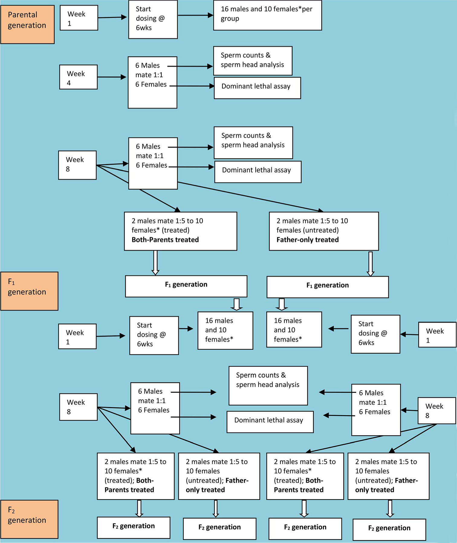
- Diagrammatic representation of the study design. *is used to indicate not all the females were dosed. Only the females that mated to simulated “both-parents treated*” were dosed. The females that mated with treated males in the “father-only treated” groups were not given ARVs.
average of both counts, multiplied by the dilution factor and divided by the chamber volume.
The second cauda epididymis was similarly minced, mixed, diluted twofold, and sieved. To assess sperm morphology, 20 µL of 1% aqueous eosin was added to 200 µL of the sperm suspension and left to stain for 30 min. Two smears were then prepared on labeled slides, air-dried, and examined microscopically for sperm head abnormalities using a standard laboratory guide.
Sperm DNA was extracted using the Qiagen DNA Mini Kit following the protocol titled “Isolation of Genomic DNA from Sperm: Protocol 2 (QA04 Jul-10).” DNA quantification was based on the method described by Georgiou et al.,[21]
which utilizes PicoGreen dye, a fluorometric probe specific to double-stranded DNA. DNA content (in µg/mL) was normalized by sperm concentration (cells/mL) to calculate DNA quantity per sperm cell in picograms.
Statistical Analysis
Data obtained were collated and analyzed using Epi Info version 3.5.3 (CDC, Atlanta) and Microsoft Excel. The Student’s t-test and analysis of variance (ANOVA) were the statistical tests applied, and a p < 0.05 was regarded as being significant. The t-test compared treatment group values to the corresponding NC group values pairwise, using a two-tailed test with different variances. ANOVA was used to compare
the different doses per drug collectively to the corresponding NC values or to each other.
RESULTS
Before therapy, the median weight (±standard deviation; range) of the parental generation was 26.0 g (±3.5; 19–38) and 16.5 g (±4.1; 10–24) for the F1 generation. The median temperature (±standard deviation; range) in the animal house for the 10-month study duration was 30.8°C (±2.2; 25.6–36.2) while the median humidity was 65% (±14; 20– 90). The results of the birth statistics have been reported elsewhere.[22]
Testicular-Body Weight (TBW) Ratios
The TBW ratios of the NC group were 0.344 and 0.366 at mid- term (week 4) and full-term (week 8), respectively. At mid- term, there was no significant difference in the TBW ratios among the NC, ZDV, and NVP groups. However, pairwise t-test analysis showed a significant decrease in TBW ratios (p = 0.038) for the NVP5 mg/kg group at full term, with a median value of 0.300 [Figure 2a]. Further comparison of the TBW ratios and body weights of individual mice in the NVP 5 mg/kg group, sampled at mid- and full-term, indicated that mice randomly sampled at full-term weighed significantly more (p = 0.002) but had significantly lower TBW ratios (p = 0.009).The F1 mice had significantly lower TBW ratios for both ZDV groups and one NVP group [Figure 2b]. Progeny from the “both-parents” treated group had lower ratios compared to the progeny from the “father-only” treated group for each drug. The F1 males were observed to generally have smaller testicular sacs, especially among the ZDV groups.
Sperm Counts
The sperm counts of the NC group were 3.6 × 10⁶/mL and 8.1 × 10⁶/mL at mid-term and full-term, respectively. At mid-term, the sperm counts of the ZDV groups were not significantly different from the NC group. However, the NVP 5 mg/kg had significantly higher sperm counts (5.6 × 10⁶/mL; p = 0.031), the NVP 50 mg/kg group had significantly lower sperm counts (2.3 × 10⁶/mL; p = 0.014), and the NVP 150 mg/kg group were not significantly different (6.0 × 10⁶/mL; p = 0.521). At mid-term, the sperm counts of one male in the NVP 50 mg/kg group and those of two males in the NVP 150 mg/kg group were too low to be determined. At full-term, the sperm counts of both the ZDV and NVP groups were all significantly lower than the NC group [Figure 2a].
The F1 generation also had significantly lower sperm counts for both ZDV groups and one NVP group [Figure 2b]. The F1 males had severely reduced sperm counts, such that the highest recorded sperm count (4.0 × 10⁶/mL) was roughly half that of the NC group. Progeny from the “both-parents” treated parental group had lower sperm counts compared to the progeny from the “father-only” treated parental group [Figure 2b].
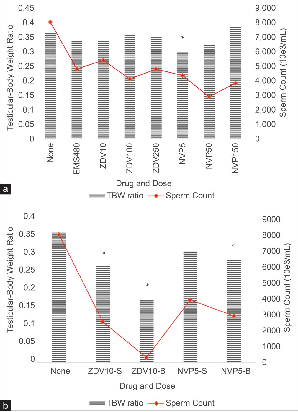
- (a) Median testicular-body weight (TBW) ratios and sperm counts for the parental (b) and F1 generation males after 8 weeks of drug administration. ZDV: Zidovudine, NVP: Nevirapine, EMS: Ethyl methyl sulfonate (1-time 480 mg/kg), –S: F1 Progeny from “father-only” treated group, –B: F1 Progeny from “both-parents” treated group. *p < 0.05 and significantly different.
Sperm Head Anomalies
The sperm head anomalies (%) recorded in the NC group were 1.89% and 2.77% at mid and full-term, respectively. Pairwise t-test analysis showed that the ZDV 250 mg/kg group (7.32%; p = 0.017) and the NVP 150 mg/kg group (19.6%; p = 0.031) had significantly higher sperm head anomalies at mid-term [Table 1]. The differences in sperm head anomalies at full term were not significantly different among any of the treatment groups. Only three and five mice were evaluated for sperm head anomalies at mid-term in the NVP 50 mg/kg and 150 mg/kg groups, respectively, due to their low sperm counts. Similarly, at full term, we could only evaluate sperm head anomalies in five mice from the ZDV 100 mg/kg group and four mice from the ZDV 250 mg/kg group due to their low sperm counts [Table 1].
| Group | Dose (mg/kg) |
Mid‑term (week 4) | Full‑term (week 8) | |||||||
| Males (n) |
Sperm anomalies (%) ±SD |
Pairwise t‑test P values | Males (n) | Sperm anomalies (%) ±SD |
Pairwise t‑test P values | |||||
| None | None | 4† | 1.89±1.78 | - | 6 | 2.77±1.87 | - | |||
| ZDV | 10 | 6 | 3.02±3.97 | 0.165 | 6 | 4.98±12.62 | 0.260 | |||
| 100 | 6 | 12.49±34.67 | 0.110 | 5* | 8.47±19.20 | 0.158 | ||||
| 250 | 6 | 7.32±5.37 | ‡0.017 | 4* | 10.57±10.53 | 0.105 | ||||
| NVP | 5 | 6 | 4.29±2.66 | 0.054 | 6 | 2.05±5.58 | 0.622 | |||
| 50 | 3* | 30.66±14.82 | 0.101 | 6 | 4.85±6.94 | 0.239 | ||||
| 150 | 5* | 19.58±12.11 | ‡0.031 | 6 | 4.38±6.50 | 0.259 | ||||
†Only four mice were randomly selected and evaluated; *Number evaluated due to low sperm counts. ‡P<0.05. SD: Standard deviation, ZDV: Zidovudine, NVP: Nevirapine; Sperm head anomalies were not determined for the positive control group. t-test analysis compared each treatment group’s values to the negative control (none)
| Drug and dose | Parental treatment | Males (n) |
Sperm anomalies±SD |
Pairwise t‑test P values |
| None | None | 6 | 2.77±1.87 | - |
| ZDV 10 mg/kg |
Father-only | 4* | 10.99±18.00 | 0.205 |
| Both-parents | 5* | 57.21±18.08 | ‡0.005 | |
| NVP 5 mg/kg |
Father-only | 2* | 24.28±18.92 | 0.357 |
| Both-parents | 6 | 14.85±29.71 | 0.091 |
‡P<0.05; *Smaller sample sizes constrained by the number of surviving pups. SD: Standard deviation, ZDV: Zidovudine, NVP: Nevirapine. t-test analysis compared each treatment group value to the negative control (none) pairwise using the double-tailed and unequal variance options in MS Excel
Only the “both-parents” ZDV 10 mg/kg-treated F1 group had significantly higher anomalies (57.2%; p = 0.005) [Table 2]. The sperm head anomalies observed in the treatment groups are shown in Figure 3.
Sperm DNA Quantity
The sperm DNA quantity for the NC group at mid-term was 52.9 µg/mL and 78.9 µg/mL at full-term. Consequently, their DNA quantity per sperm was 12.9 pg and 9.6 pg at mid- and full-term, respectively. The sperm DNA quantity of the parental generation groups was not significantly different from that of the NC group at either mid- or full- term [Figure 4]. The DNA quantity per sperm differed significantly only at full term for both the ZDV (p = 0.014) and the NVP (p = 0.0008) groups; however, these differences were not dose-dependent for either treatment group (data not shown).
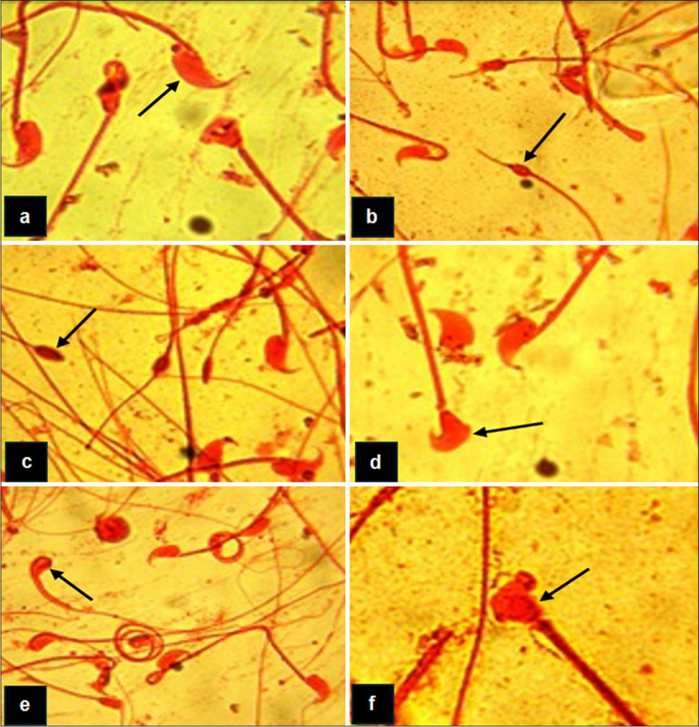
- Major sperm head morphology anomalies observed. (a) Spermatozoon (arrowed) exhibiting normal sperm head morphology, (b) Spermatozoon (arrowed) showing pin-head anomaly, (c) Spermatozoon showing a knobbed (arrowed) anomaly, (d) Spermatozoon (arrowed) exhibiting an amorphous head anomaly, (e) Spermatozoon (arrowed) illustrating the knobbed head anomaly, (f) Spermatozoon (arrowed) illustrating the hammer-head anomaly.
For the F1 males, the sperm DNA quantity was significantly smaller for both the ZDV and the NVP groups [Figure 4]. However, only the DNA quantity per sperm for the ZDV groups was significantly different (p = 0.007), with a median of 6.4 pg and 56.3 pg for the male progeny from the “father- only” and the “both-parents” treated parental groups, respectively.
Inter-Generational Comparisons
A comparison of all assayed parameters between the parental and the F1 groups showed that the ZDV 10 mg/ kg group differed significantly in all parameters. The TBW ratios, sperm counts, and DNA quantity decreased in the ZDV groups across generations, while the frequency of sperm head anomalies increased [Table 3]. The NVP 5 mg/ kg groups’ parental and F1 groups differed significantly only in sperm DNA quantity, which decreased across generations.
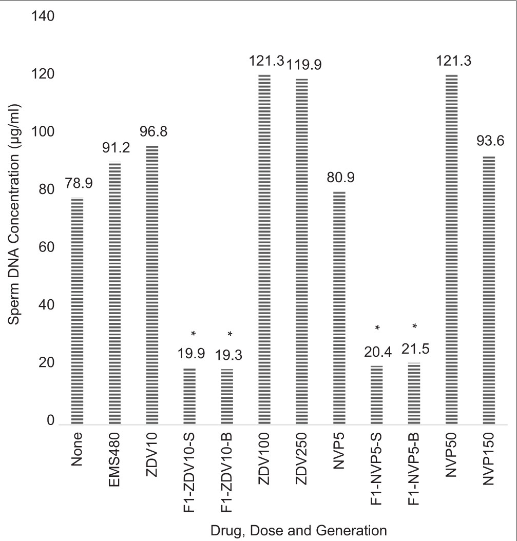
- Median sperm deoxyribonucleic acid quantity for the parental and F1 generation males after 8 weeks of drug administration. ZDV: Zidovudine, NVP: Nevirapine, EMS: Ethyl methyl sulfonate (1-time 480 mg/kg), –S: F1 Progeny from “father- only” treated group, –B: F1 Progeny from “both-parents” treated group. *p < 0.05 and significantly different.
For both the ZDV and NVP F1 groups, the TBW ratios and sperm counts were least for the “both-parent” treated groups. Three out of the four F1 groups had significantly smaller TBW ratios compared to only the NVP 5 mg/kg group among the parental generation.
DISCUSSION
The effect of administering ZDV or NVP to mice for the entire period of spermatogenesis was evaluated across two generations, evaluating various markers at different levels of the male reproductive system. We report here their effect on the male reproductive system, as their impact on fertility and birth statistics in mice has been reported.[22]
At the testicular level, the TBW ratio was determined to remove bias due to body weight and give an indication of the organ-level impact on the testes. The differences were only significant among one parental generation group, but most F1 groups had significantly lower TBW ratios. The impact of decreased testicular weight on testicular function, sperm quality, and fertility has yet to be determined. Two genetic studies involving mutant or knockout mice found that reduced testis weight resulted in lower sperm counts when specific genes were deactivated.[23,24] However, these studies also noted that fertility measured by litter size or breeding frequency and sperm DNA integrity were not adversely affected.[23,24] In the current study, only the parental group treated with 5 mg/kg of NVP showed a significantly lower TBW ratio along with decreased sperm counts at full term. This suggests that the notable decline in sperm count observed in this study may be linked to the reduction in testicular weight. Sperm counts were significantly lower among both the parental and the F1 groups, with most being less than half the sperm counts of the untreated mice. The oligospermia observed agrees with earlier reports of decreased semen volumes and sperm counts in humans.[25,26] Among the F1 mice, severe oligospermia was observed. Earlier, decreased sperm motility in humans initiating therapy was reportedly independent of the use of thymidine analogs.[15] From this mouse study, however, we can attribute a significant reduction in mice sperm counts to ZDV or NVP therapy.
| Parameter | Z10 median values | N5 median values | |||||||||
| P Z10 | F1 Z10‑S | F1 Z10‑D | ANOVA p value | P N5 | F1 N5‑S† | F1 N5‑D | ANOVA p value | ||||
| Testicular-body weight ratios | 0.34 | 0.27 | 0.18 | 0.002‡ | 0.30 | 0.31 | 0.29 | 0.693 | |||
| Sperm counts (×103/mL) | 5425 | 2570 | 320 | <0.0001‡ | 4375 | 3950 | 2945 | 0.507 | |||
| Sperm head anomaly (%) | 4.98 | 10.99 | 57.21 | 0.0002‡ | 2.05 | 24.28 | 14.85 | 0.174 | |||
| Sperm DNA quantity (µg/mL) | 96.8 | 19.9 | 19.3 | <0.0001‡ | 80.9 | 20.4 | 21.5 | <0.0001‡ | |||
| Sperm DNA per sperm (pg) | 20.6 | 6.4 | 56.4 | 0.005‡ | 18.9 | 9.3 | 7.1 | 0.011‡ | |||
†N5-S group values were from only two surviving males; ‡p < 0.05. Z10: Parental generation zidovudine 10 mg/kg group, Z10-S: F1 progeny from the zidovudine 10 mg/kg “father-only” treated group, Z10-D: F1 progeny from the zidovudine 10 mg/kg “both-parents” treated group, N5: Parental generation nevirapine 5 mg/kg group, N5-S: F1 progeny from the nevirapine 5 mg/kg “father-only” treated group, N5-D: F1 progeny from the nevirapine 5 mg/kg “both-parents” treated group, ANOVA: Analysis of variance, DNA: Deoxyribonucleic acid
There was a significant difference in sperm head anomalies between the two parental mouse groups administered the highest doses of ZDV or NVP. Among the F1 mice, there were significantly higher sperm head anomalies only for the ZDV “both-parents” treated group. Increased spermatozoa anomaly indices had been reported among HIV-infected persons, and 91% of study participants were on ART receiving two NRTIs, three or more combination therapy.[27] Our results suggest that higher sperm anomalies are attributable to ZDV therapy and agree with this previous study in humans.
The sperm DNA quantity was significantly lower only among the F1 mice. Increased mtDNA in spermatozoa has been reported among HIV-1-infected men undergoing treatment with HAART,[16] and deletions in mtDNA leading to amplification of the mtDNA have also been reported.[17] In our study, changes in total sperm DNA quantity were not significantly different among the parental mice treated with ZDV or NVP. Only among the F1 mice was the difference significant.
The intergenerational comparison showed ZDV affected molecular and morphological parameters across various levels of the male reproductive system in mice. However, NVP affected mainly the molecular parameters. The reason for this is unclear. It may be related to the competitive inhibitory nature of ZDV, which may increase its chances of incorporation into genomic DNA and, consequently, enhance the potential for DNA damage. Thus, it may have affected the development of the testes, potentially leading to oligospermia. We have earlier shown that these ARVs affect cell division by making the chromosomes sticky and separation to different poles during mitosis.[28] If replicated here, it could be associated with the wide variation in sperm DNA/sperm recorded for the F1 ZDV groups. It may have consequences for fertility and genome instability in the offspring.
Strengths and Limitations
This study utilized two generations of albino mice, making it ideal for tracking the effects of ZDV or NVP. However, the absence of genetic data on the mouse strain used is a limitation of this study. In the context of the 3Rs, this cross-generational study cannot be replaced, as the short generation time in mice provided insight into potential occurrences associated with ZDV and NVP therapy in humans. In addition, reducing the numbers used among the parental generation led to sample size challenges for the subsequent F1 studies. Hopefully, future studies could consider this and be well-powered to shed more light on the transgenerational effects. Finally, the implications and biological relevance of changes in DNA content per sperm are not clear and require further research.
CONCLUSION AND IMPLICATIONS FOR TRANSLATION
This study has shown that administering ZDV or NVP to mice for the duration of one spermatogenic cycle is associated with reduced testicular weight and sperm DNA content in the second generation, as well as reduced sperm counts in both the parental and F1 generations. Changes observed in the mouse sperm head anomalies appeared to be transient. It is possible that in humans, ZDV or NVP therapy could have the same effects produced by ZDV or NVP monotherapy in mice, especially since humans receive a triple-drug regimen as the standard of care. Furthermore, some of these effects have been reported in HIV-infected persons on ARV therapy.
It is yet to be explained why detrimental effects on sperm parameters were recorded when “both parents” were treated, and yet, they had enhanced fertility as we earlier reported,[22] in agreement with previous findings.[23,24] Further mechanistic studies are needed to elucidate these occurrences. This could guide the redesigning and/or refining of current ARVs. This study highlights the need for careful consideration in the use of ZDV and NVP for treating people living with HIV due to their potential adverse effects on male reproductive health.
Key Messages
(1) Zidovudine (ZDP) or nevirapine (NVP) administration led to reduced testicular weight across the generations. (2) Both the parental (F0) and F1 generations showed significantly reduced sperm counts following treatment. (3) ZDP or NVP administration led to the reduction of sperm DNA content and quality in the second generation. (4) Abnormalities in sperm head morphology appeared to be temporary, suggesting some degree of recovery over time.
Acknowledgments
The authors acknowledge Dr. Miriam Poirier for her mentorship, support, and donation of some of the reagents used. We thank Duncan A. Ota (Technologist at the College of Medicine, University of Lagos) for his assistance with managing the animals. We also acknowledge Prof Oni Idigbe, Dr N. Odunukwe, and Dr D. Onwujekwe, who facilitated the provision of the test drugs and animal house facilities for this research work at the Nigerian Institute of Medical Research. Extracts from this work were presented orally (TH6OP037) at the 3rd International Scientific Conference of the Nigerian Institute of Medical Research, held in Lagos, Nigeria, in November 2012.
COMPLIANCE WITH ETHICAL STANDARDS
Conflicts of Interest: The authors declare no competing interests. Financial Disclosure: Nothing to declare. Funding/Support: There was no funding for this study. Ethics Approval: The research/study was approved by the Institutional Review Board at Nigerian Institute of Medical Research, number IRB/09/094, dated September 2010. Declaration of Patient Consent: Patient consent was not required as albino mice were used for this study. The Institute’s ethics board approved the waiver at the time. Use of Artificial Intelligence (AI)‑Assisted Technology for Manuscript Preparation: The authors confirm that there was no use of artificial intelligence (AI)-assisted technology for assisting in the writing or editing of the manuscript and no images were manipulated using AI. Disclaimer: None.
References
- Toward a cure -advancing HIV/AIDs treatment modalities beyond antiretroviral therapy: A review. Medicine (Baltimore). 2024 Jul 05;103(27):e38768.
- [CrossRef] [PubMed] [Google Scholar]
- Nucleotide reverse transcriptase inhibitors: A thorough review, present status and future perspective as HIV therapeutics. Curr HIV Res. 2017 Dec 01;15(6):411-21.
- [CrossRef] [PubMed] [Google Scholar]
- Insights into HIV-1 reverse transcriptase (RT) inhibition and drug resistance from thirty years of structural studies. Viruses. 2022 May 11;14(5):1027.
- [CrossRef] [PubMed] [Google Scholar]
- HIV infection and AIDS: The present status of antiretroviral therapy. In: European handbook of dermatological treatments. Cham: Springer International Publishing; 2023 Oct 05;429-42.
- [CrossRef] [Google Scholar]
- Update on known and emergent viruses affecting human male genital tract and fertility. Basic Clin Androl. 2024 Mar 14;34(1):6.
- [CrossRef] [PubMed] [Google Scholar]
- Viral disruption of blood-testis barrier precedes testicular infection. Viruses. 2025 May 23;17(6):747.
- [CrossRef] [PubMed] [Google Scholar]
- HIV infection of the male genital tract--consequences for sexual transmission and reproduction. Int J Androl. 2010 Feb;33(1)e98-108.
- [CrossRef] [PubMed] [Google Scholar]
- Antiretroviral therapy through barriers: A prominent role for nanotechnology in HIV-1 eradication from sanctuaries. J Pharm Pharmacol. 2016;4(7)328-40.
- [CrossRef] [Google Scholar]
- Effect of in vitro exposure of first-line antiretrovirals on healthy human spermatozoa on kinematics and motility. Int Urol Nephrol. 2025 Jan 03;57(6):1715-35.
- [CrossRef] [PubMed] [Google Scholar]
- Decreased semen volume and spermatozoa motility in HIV‐1‐infected patients under antiretroviral treatment. J Androl. 2007 May 06;28(3):444-52.
- [CrossRef] [PubMed] [Google Scholar]
- Effects of antiretroviral therapy on semen quality. AIDS. 2008 Mar 12;22(5):637-42.
- [CrossRef] [PubMed] [Google Scholar]
- Investigating the effect of selected antiretroviral therapies on serum testosterone and testicular microstructure of wistar rats. Ibom Med J. 2021 Jul 01;14(3):310-8.
- [CrossRef] [Google Scholar]
- Assessment of the toxicity of different antiretroviral drugs and their combinations on sertoli and leydig cells. Drug Chem Toxicol. 2024 Nov 01;47(6):1100-8.
- [CrossRef] [PubMed] [Google Scholar]
- Molecular mechanisms underpinning the protection against antiretroviral drug- induced sperm-endocrine aberrations and testicular toxicity: A review. Reprod Toxicol. 2024 Sep:128:108629.
- [CrossRef] [PubMed] [Google Scholar]
- Dynamics of HIV viral load in blood and semen of patients under HAART: Impact of therapy in assisted reproduction procedures. AIDS. 2007 Jan 30;21(3):377-9.
- [CrossRef] [PubMed] [Google Scholar]
- Effects of antiretroviral therapy on sperm DNA integrity of HIV-1-infected men. Am J Mens Health. 2018 Nov;12(6)1835-42.
- [CrossRef] [PubMed] [Google Scholar]
- Sperm mitochondrial DNA deletions as a consequence of long term highly active antiretroviral therapy. AIDS. 2001 May 25;15(8):1061-2.
- [CrossRef] [PubMed] [Google Scholar]
- International guiding principles for biomedical research involving animals. Australas Radiol. 1985;15(1)83-7.
- [Google Scholar]
- Guideline for testing of chemicals 416, two-generation reproduction toxicity study. Paris: OECD Publishing; 2001 Jan 22. p. 1-13.
- [Google Scholar]
- Sperm head abnormalities and dominant lethal effects of formaldehyde in albino rats. Mutat Res/Genet Toxicol Environ Mutagen. 1997 Mar 17;389(2-3):141-8.
- [CrossRef] [PubMed] [Google Scholar]
- Protocol for the quantitative assessment of DNA concentration and damage (fragmentation and nicks). Nat Protoc. 2009 Feb;4(2)125-31.
- [CrossRef] [PubMed] [Google Scholar]
- Foetal loss and enhanced fertility observed in mice treated with zidovudine or nevirapine. PLoS One. 2014 Sep 18;9(9):e107899.
- [CrossRef] [PubMed] [Google Scholar]
- Disruption of the murine PIASx gene results in reduced testis weight. J Mol Endocrinol. 2005 Jun 01;34(3):645-54.
- [CrossRef] [PubMed] [Google Scholar]
- Reduced sperm count and normal fertility in male mice with targeted disruption of the ADP-ribosylation factor-like 4 (Arl4) gene. Mol Cell Biol. 2002 Apr 01;22(8):2761-8.
- [CrossRef] [PubMed] [Google Scholar]
- Semen alterations in HIV-1 infected men. Hum Reprod. 2002 Aug 01;17(8):2112-8.
- [CrossRef] [PubMed] [Google Scholar]
- The effect of human immunodeficiency virus on sperm parameters and the outcome of intrauterine insemination following sperm washing. Hum Reprod. 2004 Oct 01;19(10):2289-97.
- [CrossRef] [PubMed] [Google Scholar]
- Antiretroviral therapy effects on genetic and morphologic end points in lymphocytes and sperm of men with human immunodeficiency virus infection. J Infect Dis. 2001 Jul 15;184(2):127-35.
- [CrossRef] [PubMed] [Google Scholar]
- Exposure of allium cepa root cells to Zidovudine or Nevirapine induces cytogenotoxic changes. PLoS One. 2014 Sep 18;9(3):e90296.
- [CrossRef] [PubMed] [Google Scholar]